The Art and Science of Seeing Under the Skin
/

A ray’s skeleton is made of cartilage, which stains blue. Image by MC Gilbert, 2019.
Under the Skin is the most recent science exhibition at the Bruce Museum, on display on our renovated galleries until July 19, 2020. Under the Skin showcases some of the newest techniques being used to image biologic specimens, and the beautiful pictures that can come out of these investigations. One featured technique is clearing and staining, or diaphonization.
Cleared and stained specimens are strikingly beautiful, with colorful bones and cartilage showing through transparent flesh. The process used to make these specimens was first developed in 1927, and continues to be used by comparative anatomists, vertebrate embryologists, and anyone else with an interest in skeletal form or development.

Cleared and stained specimens at the Bruce Museum. Image by Kate Dzikiewicz.
The first stage to this multi-step process is to clean the specimen. Removal of internal organs is suggested, and animals with fur or feathers should be skinned. Next, the tissues of the animal are digested with a solution containing the enzyme trypsin. This enzyme leaves the connective protein collagen intact, creating a specimen that is ghostly translucent, but will not fall apart.
The specimen is next bathed in a solution containing the dye Alizarin red, which stains bones red. Cartilage is likewise dyed blue using Alcian blue. When the treated specimen is placed in glycerin, something amazing happens: The cloudy connective tissues seemingly disappear and become transparent, revealing the colored skeleton inside. This is because glycerin has the same refractive index as collagen, allowing light to pass through unimpeded. Now, the skeleton of the organism appears suspended in perfect articulation and precise color, and is ready for study.
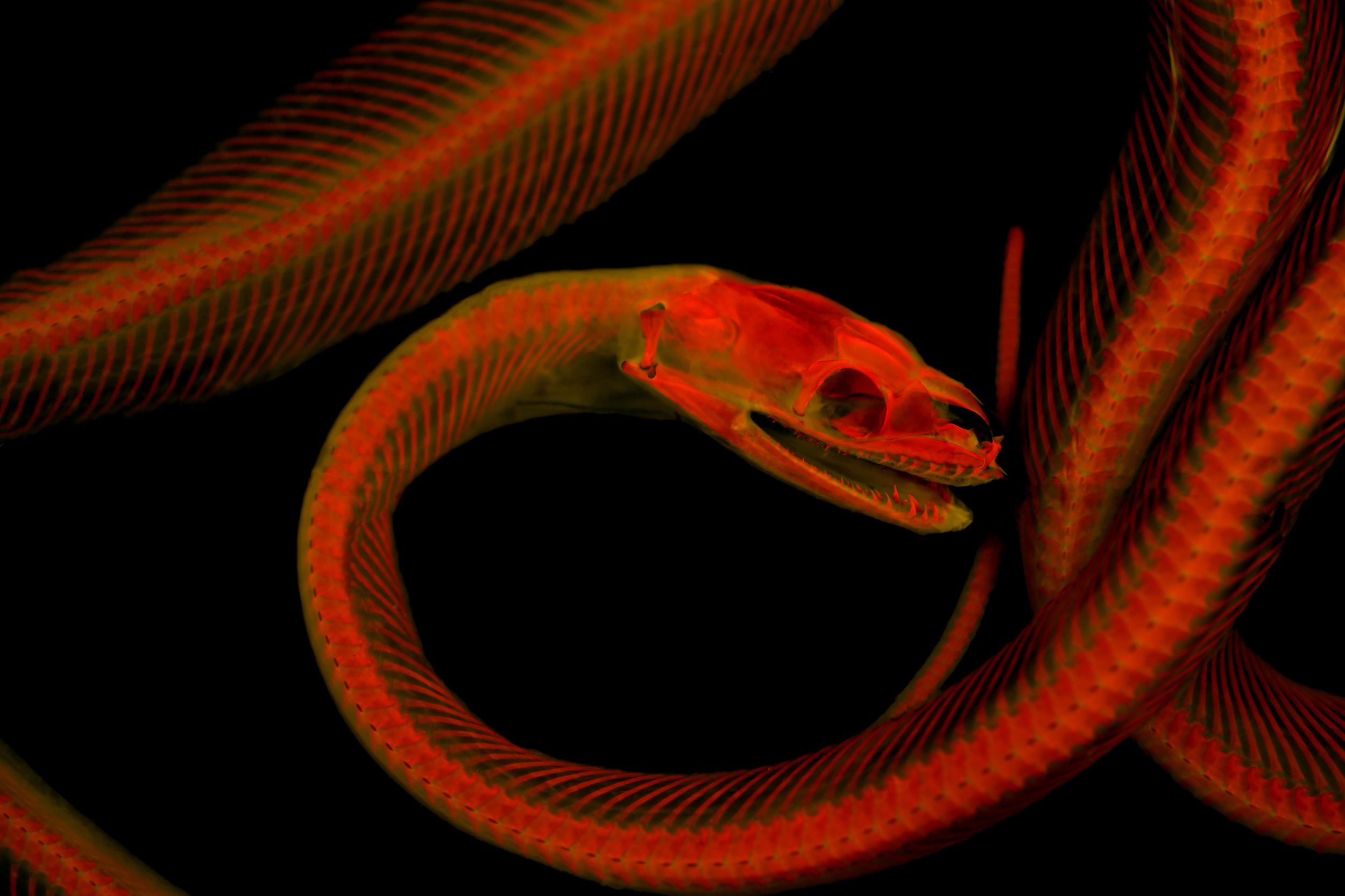
Macklot’s Python (Liasis mackloti). Cleared and stained specimen under fluorescent light. Image courtesy Dr. Matt Girard and Dr. Leo Smith.
Why is this technique still in use when we have many more high-tech ways of imaging the interior of the body, such as radiographs or CT scans? As it turns out, there are many benefits to diaphonization. One aspect is cost. Diaphonization requires very little in the way of specialized equipment, while an x-ray machine can cost tens to hundreds of thousands of dollars. Also, even if a scientist has a high tech scanner, diaphonization still has perks. Cartilage is of much lower density than bone and is hard to detect with many internal imaging methods, but is easy to see when an organism is cleared and stained.
Recently, scientists discovered another perk: Alizarin red fluoresces under certain wavelengths of light. The fluorescing bone illuminates small anatomical details and makes them easier to study and photograph. In addition to the scientific utility of this feature, the images themselves can be stunning, looking equally at home in a scientific journal or on the wall of a tattoo parlor. Many images of fluorescing cleared and stained specimens can be seen in the Under the Skin exhibition, as well as the specimens themselves. Want to learn more about this topic and many others? It’s time to visit the Bruce!
- Kate Dzikiewicz, Science Curatorial Associate and Seaside Center Manager